Opening Times
- Mon 09:00 - 17:00
- Tues 09:00 - 17:00
- Wed 09:00 - 17:00
- Thurs 09:00 - 17:00
- Fri 09:00 - 17:00
"Serving my time in the Armed Forces requires a lot of sacrifice, commitment and dedication. Fitness is my everyday life. You'll never know when you are called out on deployment or exercise out in the field and at times I feel the stress and body aches upon returning out from the field. It's all part of everyday life in the Army. I always make sure I allow myself a good rest after work and I'm very fortunate and lucky to see Kay in the clinic!" Mr I.T
Address
8 Maristow Street
Westbury
Wiltshire
BA13 3DN
01373 865533
Associations


Diagnostic Ultrasound Service
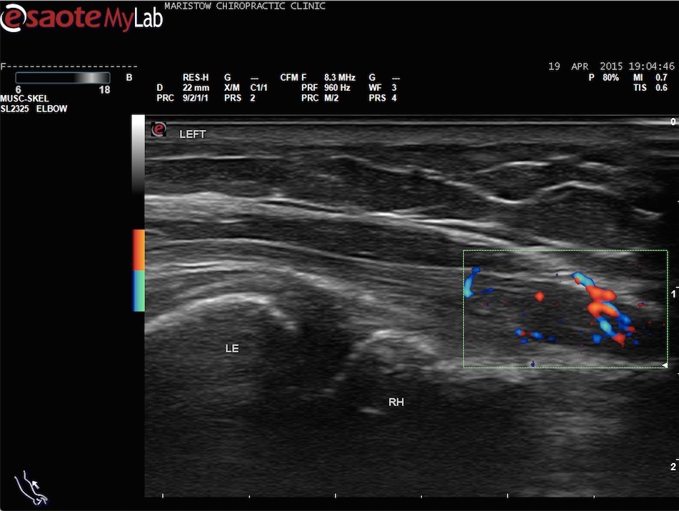
An ultrasound scanner uses high frequency sound waves which can’t be heard by the human ear. An instrument passed over the skin uses these waves to create an image of internal structures of the body. Ultrasound is particularly useful for diagnosing muscle, tendon and ligament injuries. Many people are familiar with this technology for imaging unborn babies.
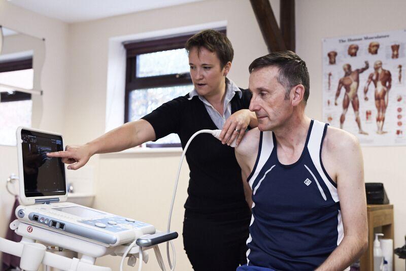
In 2016 Kay Pearce completed a Post Graduate Diploma in Medical Ultrasound (PgDip), which is the Consortium for the Accreditation of Sonographic Education (CASE) accredited course, recognised by the British Medical Ultrasound Society (BMUS). Having this qualification and access to an ultrasound machine enables us to more accurately diagnose a patients injuries, and more importantly, provide a specific treatment plan and prognosis.
We can also quickly recognise complaints that are not suitable for manual therapy so can save patients time and money on ineffective treatments.
How does it work?
In nature, the same principles are employed by bats as sonar. In technology, ships and angling boats use this principle to detect shoals of fish or obstacles in the water. A transducer pressed against the skin directs a stream of inaudible, high-frequency sound waves into the body. These sound waves bounce against the internal structures within the body and the transducer records tiny changes in the sound's pitch and direction. The echo wave is instantly measured, recorded and depicted as a image on a screen, showing us how far away a structure is, its shape, size, and if it’s fluid, solid or something in between.
What are the benefits of ultrasound?
With both MRI and X-ray the patient has to remain very still, but with ultrasound we can move the patient to see how the soft tissue changes under stress. For example subtle tendon tears may not show unless we stretch them and swelling may not present itself unless we flex a joint. With ultrasound systems you can watch the soft tissues moving or getting stuck! We can even scan whilst the patient recreates the movement that causes them pain.
Other advantages:
• Ultrasound scanning is usually painless and non-invasive.
• Ultrasound does not use ionising radiation.
- Ultrasound is not affected by cardiac pacemakers, implants or fragments within the body.
- Ultrasound can also be an excellent alternative to MRI for claustrophobic patients.
- Ultrasound may actually have advantages over MRI in seeing tendon structure, which is better appreciated by ultrasound than MRI.
- The images can be viewed and interpreted immediately.
Are there any risks?
For standard diagnostic ultrasound there are no known harmful effects on humans (The American Institute of Ultrasound in Medicine 2012).
The skin over the area to be examined needs to be unbroken. If you have any allergies which may be affected by the gel, please advise at point of booking.
What are the limitations of Ultrasound Imaging of the Musculoskeletal System?
Ultrasound waves have difficulty penetrating bone. Therefore we can only see the outer surface of bony structures and not what lies within and beyond. For visualising bone or internal structure of certain joints MRI or X-ray may be more suitable. It is important that you are directed towards the most appropriate form of imaging. This is one of the reasons we ask you to have a consultation with one of our chiropractors, or a referral from another MSK practitioner, prior to an ultrasound scan.
Costs:
£92 for external referrals.
Discounts may apply for registered patients, please contact us for details.
Referring musculoskeletal practitioners please use the form below prior to referring a patient for an ultrasound scan: